For as long as neuroscience has studied voluntary movement, the answer to 'how do humans control their hands?' has pointed in one direction: the cortex. The brain's large, wrinkled outer layer — the command centre for conscious thought — sends signals directly down the spinal cord to the muscles of the arm and hand. Clean, direct, well-understood.
Except, as a new UC Riverside-led study published March 12, 2026 in the *Proceedings of the National Academy of Sciences* reveals, that picture has been missing something important.
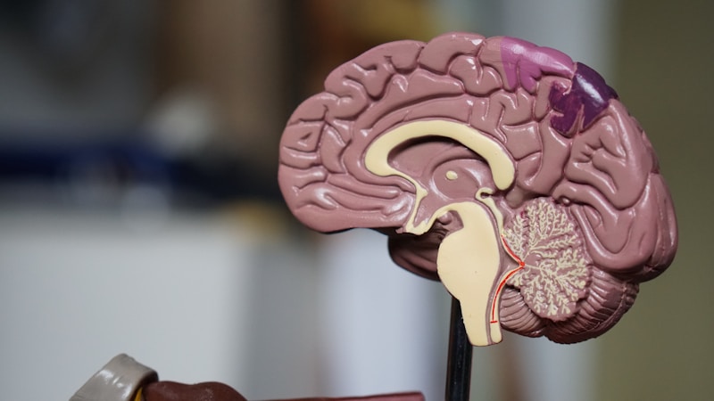
Between the cortex and the spinal cord lies the **brainstem** — a narrow stalk of tissue that regulates breathing, heart rate, posture, and balance, and that scientists had long assumed played a limited role in fine, voluntary hand movements. The UC Riverside team found that assumption was wrong.
**The Discovery**
Led by **Shahab Vahdat**, an assistant professor of bioengineering at UC Riverside, the research team used functional magnetic resonance imaging (fMRI) to examine brain and brainstem activity during controlled hand movements in both mice and humans.
The results revealed a **multi-stage pathway** that had been largely overlooked. Signals controlling voluntary hand movements don't only travel directly from the cortex to the spinal cord. They also route through two relay centres in the medulla — the lowest portion of the brainstem, which sits just above the cervical spinal cord — before reaching the muscles of the hand and arm.
In mice, the team trained animals to press a small lever with varying force while recording neural activity across the brain and brainstem. In humans, volunteers performed a similar gripping task in the MRI scanner. In both species, the medulla lit up in a pattern consistent with an active relay role — not merely a passive conduit.
'For a long time, we thought fine hand movements in humans were controlled almost entirely by the cortex,' said Professor Vahdat. 'What we are observing is that evolutionarily older brainstem structures also play an important role.'
**Why This Changes How We Understand Movement**
The hands are extraordinary instruments. Human fine motor control — the ability to grasp, manipulate, thread a needle, play a piano, type at speed, unscrew a bottle — is among the most complex motor behaviour in the animal kingdom. Explaining how the brain coordinates this has been one of neuroscience's central challenges.
The direct corticospinal pathway — cortex to spinal cord — is well established and clearly important. But the new findings suggest a **parallel system** operating alongside it. The brainstem relay centres in the medulla appear to process and modulate the descending movement signals in ways that may be critical for the precision of the final output.
The specific medullary regions activated — identified through detailed anatomical mapping — act as crossroads where signals from the cortex are integrated with information about posture, balance, and body position. This integration may be essential for the context-sensitivity of skilled hand movements: grasping differently depending on the object's weight and fragility, for example.
**The Stroke Connection**
The finding carries significant therapeutic implications, particularly for stroke recovery.
Stroke — which kills or disables approximately 15 million people globally each year — frequently damages the cortex or the direct corticospinal pathways that descend from it. The result is partial or complete loss of hand and arm function: the inability to grip, write, button a shirt, make a cup of tea. It's one of the most debilitating and common consequences of stroke.
Current rehabilitation approaches focus on rebuilding the direct corticospinal pathway through intensive practice and neurostimulation. But many stroke survivors reach a plateau where further recovery stalls, even with continued therapy.
If the brainstem pathway discovered by the UCR team is intact after a stroke that damages the cortex — or if it can be strengthened or recruited as a compensatory route — it may offer a new therapeutic target. Stimulating the medullary relay centres, or designing rehabilitation protocols that specifically engage this pathway, could potentially unlock recovery that direct corticospinal rehabilitation cannot achieve.
'By mapping this pathway, researchers say the work could help guide new therapies aimed at restoring hand and arm function after stroke or other neurological injuries,' the team stated at publication.
**An Evolutionarily Ancient Circuit**
Another layer of significance: the brainstem is one of the oldest parts of the vertebrate nervous system, evolutionarily. The finding that it participates meaningfully in sophisticated voluntary hand movements suggests that the capacity for fine motor control evolved, in part, by building on ancient neural infrastructure — rather than replacing it with purely cortical control.
This is consistent with broader trends in neuroscience that have found the brain's 'primitive' regions — the brainstem, cerebellum, basal ganglia — are far more involved in sophisticated cognitive and motor tasks than the textbooks once assumed.
The hands we use to build, to make music, to hold each other's hands — their extraordinary capabilities may have roots older than the cortex we credit for them. 🧠
*Sources: UC Riverside News (news.ucr.edu, March 12, 2026) · Proceedings of the National Academy of Sciences (PNAS) · Professor Shahab Vahdat, UCR Department of Bioengineering*